Exploring Decompression Bubbles Using Advanced Ultrasound Techniques

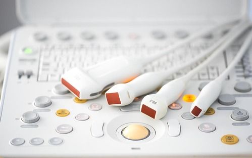
Venous gas emboli (VGE) are bubbles that can appear in the blood after a dive due to decompression. These bubbles are detectable using ultrasound imaging and provide a measure of decompression stress. However, how and where VGE form is still not fully understood. There are also unexplained differences in VGE size and quantity and decompression sickness (DCS) risk among and within individuals. Microbubbles are hypothesized to be precursors to VGE, and the ability to detect and measure these microbubbles can provide a new tool in studying decompression physiology and DCS risk. Advanced ultrasound imaging techniques are being developed at the University of North Carolina (UNC) for detecting these microbubbles and differentiating them from VGE.
Bubbles known as venous gas emboli (VGE) can appear in the venous circulation during ascent due to the offgassing of nitrogen from the tissues. Normally the lungs are able to effectively filter them from the bloodstream. Bubbles are commonly measured in diving research using ultrasound imaging of the heart, where they can be seen circulating in the venous heart chambers. Although there is a correlation between bubble load and DCS risk, VGE do not provide a direct measure of DCS.
VGE are hypothesized to grow from tiny microbubbles (smaller than a red blood cell) that are naturally present in the body. Several studies have shown using ultrasound that these microbubbles are produced during diving and after exercise. In medical ultrasound imaging, engineered microbubbles are commonly used as a vascular contrast agent (these are injected in the bloodstream). Advanced ultrasound imaging techniques have been developed to image these microbubbles specifically. Using these ultrasound imaging techniques, it has been shown that ultrasound signals increase post-dive and have different dynamics to VGE detected with normal ultrasound imaging.
At UNC, researchers are repurposing these imaging techniques to selectively detect decompression microbubbles using state-of-the-art ultrasound scanners. Furthermore, they are working on bringing this technology from the lab bench to the field where divers can be studied.
This study is a collaborative effort between the University of North Carolina, Chapel Hill and DAN, and was initiated with funding through the DAN/R.W. Hamilton Dive Medicine Research Grant in 2017.
Posted in Research
Posted in Ultrsound, Venous gas emboli, VGE, Gas emboli, Microbubbles, Bubble detection
Posted in Ultrsound, Venous gas emboli, VGE, Gas emboli, Microbubbles, Bubble detection
Categories
2025
2024
February
March
April
May
October
My name is Rosanne… DAN was there for me?My name is Pam… DAN was there for me?My name is Nadia… DAN was there for me?My name is Morgan… DAN was there for me?My name is Mark… DAN was there for me?My name is Julika… DAN was there for me?My name is James Lewis… DAN was there for me?My name is Jack… DAN was there for me?My name is Mrs. Du Toit… DAN was there for me?My name is Sean… DAN was there for me?My name is Clayton… DAN was there for me?My name is Claire… DAN was there for me?My name is Lauren… DAN was there for me?My name is Amos… DAN was there for me?My name is Kelly… DAN was there for me?Get to Know DAN Instructor: Mauro JijeGet to know DAN Instructor: Sinda da GraçaGet to know DAN Instructor: JP BarnardGet to know DAN instructor: Gregory DriesselGet to know DAN instructor Trainer: Christo van JaarsveldGet to Know DAN Instructor: Beto Vambiane
November
Get to know DAN Instructor: Dylan BowlesGet to know DAN instructor: Ryan CapazorioGet to know DAN Instructor: Tyrone LubbeGet to know DAN Instructor: Caitlyn MonahanScience Saves SharksSafety AngelsDiving Anilao with Adam SokolskiUnderstanding Dive Equipment RegulationsDiving With A PFOUnderwater NavigationFinding My PassionDiving Deep with DSLRDebunking Freediving MythsImmersion Pulmonary OedemaSwimmer's EarMEMBER PROFILE: RAY DALIOAdventure Auntie: Yvette OosthuizenClean Our OceansWhat to Look for in a Dive Boat
2023
January
March
Terrific Freedive ModeKaboom!....The Big Oxygen Safety IssueScuba Nudi ClothingThe Benefits of Being BaldDive into Freedive InstructionCape Marine Research and Diver DevelopmentThe Inhaca Ocean Alliance.“LIGHTS, Film, Action!”Demo DiversSpecial Forces DiverWhat Dive Computers Don\'t Know | PART 2Toughing It Out Is Dangerous
April
